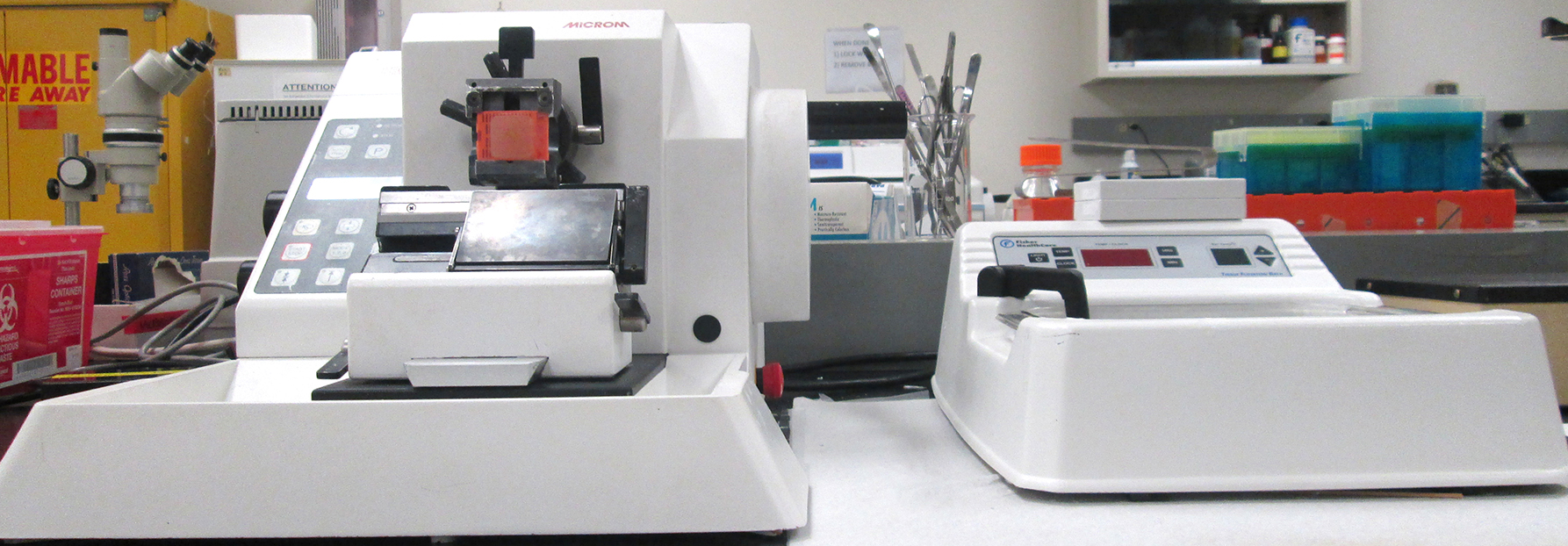
Microm HM 360 Microtome
Brief Description
Microm HM 360 rotary microtome is a heavy-duty, automated, multi-purpose instrument used for paraffin embedded tissues, hard specimens, semi-this techniques and plastic sectioning for biology, medicine and industry applications. The knife moves toward the specimen with section thickness range from 0.25 to 60 microns. It has the ability to program a series of sections for a range of specific applications. The specimen and knife edge can be quickly adjusted using the The electronically controlled motor drive and ergonomic characteristics makes the motorized coarse feed system. This microtome features a trimming function which allows for fine adjustments during the first cuts, from 5 to 500 microns, resulting in larger section thickness when trimming specimen blocks. All adjustments and settings are conveniently displayed on the digital operating panel allowing user-friendly operation. The operating panel also offers users the ability to program and store cutting sequences for trimming and sectioning series as well as, the parameters of sections thickness, number of sections and cutting speed. The Microm HM 360 rotary mictotome is ideal for producing and maintaining reproducible precision during sectioning, as well as superior section quality for every field of application.
Click here to learn more about the Microm HM 360 Rotary Microtome
Applications
Sectioning is accomplished by using a cutting apparatus called a microtome. A microtome is a instrument for holding and advancing the specimen while moving it past the cutting edge of a sharp knife. The tissue sample which has been embedded or surrounded by hard paraffin is placed in the chuck of a microtome where it will drive a knife across the surface of the paraffin cube and produce a series of thin sections with very precise thickness. The objective is to produce a continuous "ribbon" of sections adhering to one another by their leading and trailing edges. The thickness of the sections can be preset, and a thickness between 5 - 10 μm is optimal for viewing with a light microscope. Sections are cut, then removed from the blade and floated on a water bath. The sections can then be mounted on individual microscope slides which are then placed on slide warming trays to help flatten the sections out.
Preparation and mounting of the embedded tissue block on the microtome is very important to successful sectioning. Sectioning is the most difficult part of the tissue preparation process.

